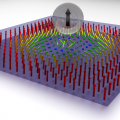
 A new technique for imaging spin properties at the nanoscale, Scanned Spin‐Precession Microscopy, works by incorporating a scannable micromagnetic tip in conjunction with any of a variety of established spin detection tools—electrical or optical, and improves upon their limited or non‐existent imaging capabilities. The magnetic field gradient from the probe directly selects spins from certain regions of the sample for study. The technique can achieve high resolution, beyond the optical diffraction limit, governed by the field gradient strength in a manner analogous to MRI.
This new tool should help in further understanding the microscopic details relevant to spin and its transport and will be an asset to researchers in spintronics, especially in the study of technologically important materials such as silicon and graphene that have been challenging to investigate with current tools. The new technique, pioneered by a collaborative team of experimentalists and theorists from OSU and Texas A&M, is to be highlighted as an Editor’s Suggestion [1] in Physical Review Letters.
The full text of the article is available here and will be published in Physical Review Letters on 13 September 2013.
[1] http://prl.aps.org/toc/PRL/v111/i11#highlighted‐articles