Currently the treatment of neurological
and neuromuscular disorders is a significant problem in medicine (e.g. spinal
cord injuries). Although great strides have been made recently in
understanding the fundamental aspects of neuroscience, the development of
advanced materials and devices for interrogating neural function lags behind
neuroscience research (deep brain stimulation invented in the 1930s is the
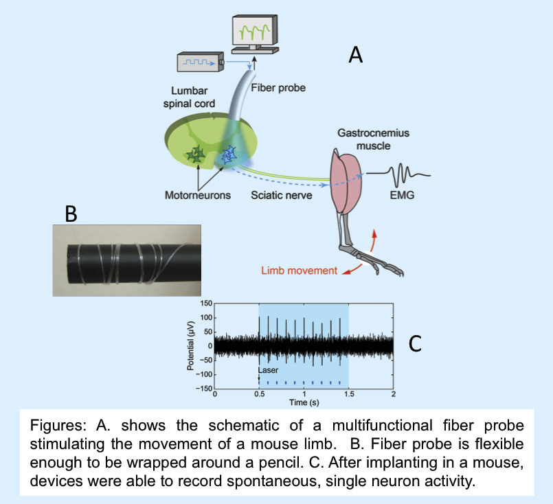
current standard). MIT MRSEC researchers have developed flexible,
multifunctional implantable probes that make it possible to simultaneously
stimulate and record single cell neural activity, with the option of delivering
therapeutic agents through the probe. Using these new probes, they report for
the first time, the direct control of leg movement via optical spinal cord
stimulation in a live mammal (see figure). The new tools provided by this
technology may someday help the millions of people suffering from disrupted
neural activity (Parkinson’s disease, depression, paralysis).
Flexible neural
probes were fabricated using a thermal drawing process. In this process, a
macroscale template of the final device is reduced in size by applying
controlled heat and stress. Different structures were fabricated:
multielectrode probes (7 electrodes, 85 μm in diameter) in which polyetherimide
(PEI) surrounds tin (Sn) electrodes, and multifunctional probes with a
waveguide (polycarbonate (PC) core and cyclyc olefin copolymer (COC) cladding),
polymer conductive polyethylene electrodes (CPE) and hollow channels for fluid
delivery. After implanting these devices in mouse they were able to record
spontaneous, single neuron activity. Multifunctional probes were also used to
excite neurons containing the light-sensitive ion channel channelrhodopsin1
using the waveguide while the neuronal activity was modulated by the injection
of drugs through the fluid delivery channels, and to record the neural activity
throughout this experiment.