Outcomes: Penn MRSEC researcher Janmey and colleagues showed that mechanical shear strain on fibrin gels — the protein scaffolding that holds a blood clot together — speeds up both of fibrin's natural remodeling enzymes: Factor XIIIa, which covalently crosslinks the fibers, and plasmin, which proteolytically degrades them.
Impacts and Benefits: Fibrin acts as a mechanoresponsive substrate whose biochemistry is set by its mechanical state. The result has direct bearing on clot stability under flow, wound healing, and the design of strain-adaptive biomaterials, and it provides a biological precedent for the IRG-1 program in materials that learn from local physical conditions.
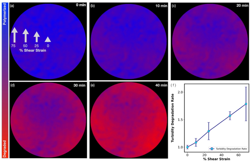
Explanation: Blood clots form under the mechanical stresses of flow, gravity, and muscle contraction, and they are remodeled afterward by enzymes that either reinforce the fibrin network (Factor XIIIa) or break it down (plasmin). Whether the mechanical state of an individual fiber changes how those enzymes act on it was unclear. The team polymerized fibrin gels in a rheometer, applied volume-conserving shear strain, and measured the consequences using shear rheology, turbidity imaging, confocal microscopy, and gel electrophoresis. Strain aligned and stretched the fibers; in the strained gels the γ-γ covalent dimers produced by Factor XIIIa accumulated more rapidly, and plasmin-driven loss of the storage modulus accelerated by nearly fourfold at 75% strain. Turbidity maps of gels under radially graded shear showed degradation propagating fastest at the outer edge, where strain is highest, and slowest at the strain-free center. Both enzymes therefore react with fibrin in proportion to the local mechanical state of the fiber, identifying fibrin as a substrate whose biochemical fate is set by the forces it carries.
Mechanical strain modulates enzymatic remodeling of fibrin networks
UPENN Materials Research Science and Engineering Centers
The LRSM at UPENN is a center of excellence for materials research and education. It facilitates collaboration between researchers from different disciplines including physics, chemistry, engineering, and biology to advance transformative scientific projects and solve societal challenges.