R.M. Suter /CMU MRSEC, Carnegie Mellon University, NSF DMR- 0520425
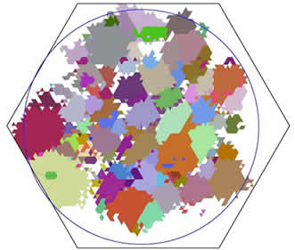 Researchers in the CMU MRSEC, together with scientists at the advanced photon source, have developed a non-destructive method to visualize the arrangement and orientation of individual crystals within a solid material. High energy X-rays from a synchrotron light source penetrate the material and interact with the crystals in their path. The pattern of transmitted X-rays that emerges from the material is then analyzed by custom software to determine the internal structure of the material. Because the complete X-ray/crystal interaction is modeled, this technique yields far more data than is contained in conventional radiograms. This new tool allows scientists to see within opaque materials with unprecedented detail and, therefore, allows the visualization of a wide range of previously hidden processes, such as crack formation in structural materials
The figure shows a two-dimensional slice of the microstructure inside of an aluminum
wire, 1 mm in diameter (the blue circle). Each color corresponds to a different crystal
orientation.
This research was conducted by: R.M. Suter, C. Xiao, D. Hennessy, and U. Lienertb
Carnegie Mellon, Department of Physics and Advanced Photon Source
Read more: http://www.andrew.cmu.edu/user/suter/3dxdm/3dxdm.html
Download .pdf file